- Arms
- Arms subequal in length, tips not attenuate.
- Arms without keels.
- Buccal connectives attach to the ventral margins of arms IV.
- Suckers with "smooth" inner rings.
- Suckers in 3 series over most of arm length.
- Arms with low protective membranes without obvious trabeculae.
- Arm suckers with inner rings that appear smooth.
- Tentacles
- Aboral side of stalk with deep groove (sulcus) running along its proximal third (see lower portion of photograph on near left and arrow in lower right photograph).
- Base of tentacle much thicker than adjacent arms.
- Club without keel, carpal locking-apparatus or terminal pad.
- Club suckers on distal 2/3 to 3/4 of tentacle.
- Club suckers with inner rings that appear smooth.
- Head
- Eye small
- Eye opening contracted into raised circular ridge.
- Large oval dorsal funnel organ.
- Funnel locking-apparatus with deep, oval depression.
- Viscera
- Anus lacks anal flaps.
- Funnel organ with large, oval dorsal pad (within the funnel in the photograph and anterior to the anus).
- Ink sac apparently absent. An ink sac could not be seen but the squid was not dissected further to look for it.
- Photophores
- Photophores absent.
- Pigmentation
- Tentacle stalks (ca. 25-35% of club length) heavily pigmented with purple-brown color (see photographs above).
- Covering of viscera (see photograph above), buccal membrane and oral surfaces of arms (see photograph on previous page) with dark purple-brown pigment.
- Measurements
- Mantle length - 25 mm (label in jar indicates 40 mm ML)
- Fin length - 9 mm
- Fin width - 18 mm
- Head length - 8 mm
- Head width - 9 mm
- Eye diameter - 2.5 mm
- Arm I (left) - 8 mm, 53 suckers
- Arm I (right) -9 mm, 45+
- Arm II (left) -9 mm, 50 suckers
- Arm II (right) - 8 mm, 46+
- Arm III (left) - 8 mm, 48 suckers
- Arm III (right) - 8 mm, 44 suckers
- Arm IV (left) - 9 mm, 53 suckers
- Arm IV (right) - 9 mm, 50 suckers
- Tentacle length (left) - 35 mm
- Tentacle length (right) - 37 mm
- Tentacle width near base - 3.8 mm
- Arms III and IV width near base - 1.9 mm
All measurements are of the holotype. The difference between the ML on the label and the present measurement indicates that considerable shrinkage has occurred in preservation.

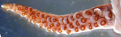
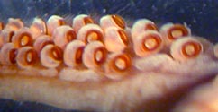
Figure. Oral view of an arm of P. sulcus, holotype. Left - Arrangement of suckers. Right - Arm base showing low protective membrane. Photographs by R. Young.
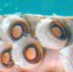
Figure. Oral view of arm suckers of P. sulcus, holotype, that appear smooth under the dissecting microscope. Photograph by R. Young.
Scanning electron microscope photographs of the suckers can be found here.
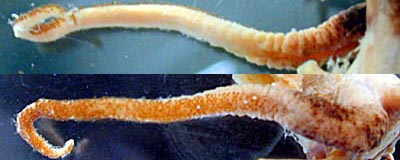
Figure. Overview of the tentacles of P. sulcus. Top - Aboral view. Bottom - Oral view. Note the pigmented base and the restriction of the club to about half the tentacle circumference. Photographs by R. Young.


Figure. Club suckers of P. sulcus, holotype. Left - Oral view of the mid-region of the club showing arrangement of suckers. Right - Higher magnification of the "smooth" suckers. Photographs by R. Younng.
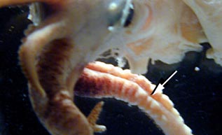
Figure. Side view of ventral portion of head and tentacle bases of P. sulcus, holotype. Arrow points to the deep aboral groove in the tentacle base. Photograph by R. Young.
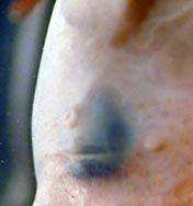
Figure. Ventrolateral view of the head of P. sulcus showing the small eye opening. Note the papilla-like olfactory organ to the right of the eye. Photograph by R. Young.

Figure. Frontal view of the funnel locking-apparatus of P. sulcus, holotype. Photograph by R. Young.
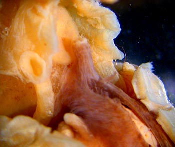
Figure. Ventrolateral view of anterior portion of mantle cavity of P. sulcus. Note the visceral pigmentation.
Comments
The gladius and beaks were not removed from the squid for examination.


