Oh my goodness! Unless you are a Tree of Life developer,
you really shouldn't be here. This page is part of our beta test site, where we
develop new features for the ToL, often messing up a thing or two in the
process. Please visit the official version of this page, which is available
here.

Click on an image to view larger version & data in a new window

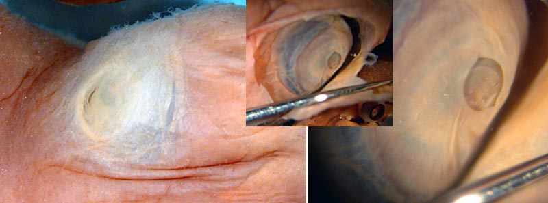
Figure. Left - A dorso-lateral view of the eye of P. sloani and the translucent "pseudocornea" and the small, crescent-shaped eye opening. Middle - A ventro-lateral view of the eye with the eyelid cut away. Right - A close-up view of the same picture. Note the iris covering much of the lens.

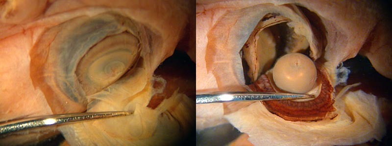
Figure. P. sloani eye dissection. Left - The iris has been removed removed, exposing the lens and the ciliary body. Right - The eyeball has been cut open and the lens and ciliary body folded over.
Comments
The eye, although somewhat squashed during capture and fixation, appears to have a normal construction although the ciliary body does not attach at the lens equator. Dissection is of the paratype. Photographs by R. Young.
About This Page
Richard E. Young

University of Hawaii, Honolulu, HI, USA
Michael Vecchione

National Museum of Natural History, Washington, D. C. , USA
Page copyright © 2003
Richard E. Young
and
Michael Vecchione
 Page: Tree of Life
Promachoteuthis sloani: Eye Structure
Authored by
Richard E. Young and Michael Vecchione.
The TEXT of this page is licensed under the
Creative Commons Attribution-NonCommercial License - Version 3.0. Note that images and other media
featured on this page are each governed by their own license, and they may or may not be available
for reuse. Click on an image or a media link to access the media data window, which provides the
relevant licensing information. For the general terms and conditions of ToL material reuse and
redistribution, please see the Tree of Life Copyright
Policies.


 Go to quick links
Go to quick search
Go to navigation for this section of the ToL site
Go to detailed links for the ToL site