Oh my goodness! Unless you are a Tree of Life developer,
you really shouldn't be here. This page is part of our beta test site, where we
develop new features for the ToL, often messing up a thing or two in the
process. Please visit the official version of this page, which is available
here.
Structure of the Amphilinidea Juvenile and Adult
Klaus Rohde
Adult worms are dorso-ventrally flattened and not divided into a strobila
(chain) of proglottids ("segments") as in the eucestodes.
They are hermaphroditic, each worm containing one set of male and one
set of female reproductive organs (Figs. 1 and 2) (Dubinina, 1982).

Click on an image to view larger version & data in a new window
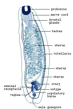
Figure 1. Juvenile Amphilina foliacea.
Note uterine and vaginal openings at anterior end, and male gonopore
at posterior end (redrawn from Dubinina, 1982).

Click on an image to view larger version & data in a new window
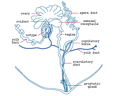
Figure 2. Adult Amphilina
foliacea with vaginal opening and male
gonopore, and various reproductive organs and ducts (redrawn from
Dubinina, 1982).
The uterine opening is located at the anterior end and near it are
penetration glands which produce histolytic secretion. The main excretory
ducts are arranged in various ways. For example, in Amphilina
foliacea they form a network (Fig.3A), in Gephyrolina
paragonopora two longitudinal ducts are connected by transverse
ones (Fig.3B).

Click on an image to view larger version & data in a new window
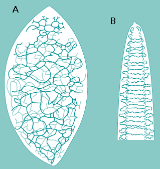
Figure 3. Excretory ducts of Amphilina
foliacea (A) and Gephyrolina
paragonopora (B) (redrawn from Dubinina,
1982).
Sensory receptors are of several types (Fig.4), although not as many
have been demonstrated as in the larva, possibly because of the large
size of the worms which makes it difficult to find all the receptor
types.

Click on an image to view larger version & data in a new window
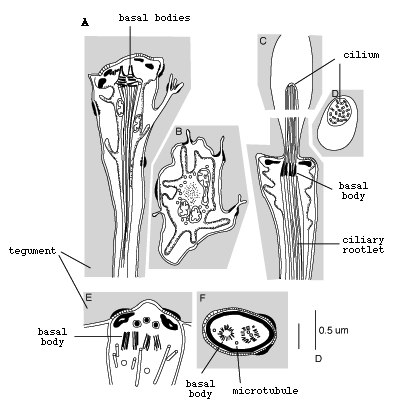
Figure 4. Sensory receptors of juvenile Austramphilina
elongata based
on electron-microscopic studies (redrawn from Rohde and Watson, 1990a).
During vitellogenesis (formation of yolk), cytoplasm of small yolk
cells gradually increases in volume, and many lipid droplets and shell
protein granules are formed which contribute to the formation of the
egg shell ( Fig.5).

Click on an image to view larger version & data in a new window
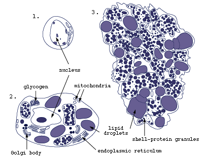
Figure 5. Vitellogenesis (yolk formation) in Austramphilina
elongata.
1. young yolk cell, 2. maturing yolk cell, 3. yolk. Note the formation
of lipid droplets and shell-protein granules (redrawn from Xylander,
1988).
Spermiogenesis is as in other parasitic platyhelminths, i.e., spermatids
form clusters connected by a central cytophore, spermatids are transformed
to sperm by elongation, and two flagella grow out which fuse with the
sperm body in a proximo-distal direction, i.e., beginning near their
base. Finally, sperm separate from the cluster (Rohde and Watson, 1986a).
References
Bazitov, A., Lyapkalo, A. and Yukhimenko (1979). [Spermatogenesis of Amphilina japonica (Goto et Ishii, 1936) (Amphilinidea)]. Vestnik Zoologi (Kiew) 1, 50-55 (In Russian).
Coil, W. H. (1987). The oogenotop of Amphilina bipunctata (Cestodaria). Parasitology Research 73, 75-79.
Coil, W. H. (1991). Platyhelminthes: Cestoidea. In "Microscopic Anatomy of Invertebrates, Vol. 3: Platyhelminthes and Nemertinea", (F. W. Harrison and B. J. Bogitsh, eds). Wiley-Liss, New York, pp. 211-283.
Dubinina, M.N. (1982). Parasitic worms of the class Amphilinida (Platyhelminthes). "Nauka", Leningrad (in Russian). (Older references therein).
Rohde, K. (1994). The minor groups of parasitic Platyhelminthes. Advances in Parasitology 33, 145- 234.
Rohde, K. and Watson, N. (1986a). Ultrastructure of spermatogenesis and sperm of Austramphilina elongata (Platyhelminthes, Amphilinidea). Journal of Submicroscopic Cytology 18, 361-374.
Rohde, K. and Watson, N. (1986b). Ultrastructure of the sperm ducts of Austramphilina elongata (Platyhelminthes, Amphilinidea). Zoologischer Anzeiger 217, 23-30.
Rohde, K. and Watson, N. (1989). Ultrastructural studies of larval and juvenile Austramphilina elongata (Platyhelminthes, Amphilinidea); penetration into, and early development in the intermediate host, Cherax destructor. International Journal for Parasitology 19, 529-538.
Rohde, K. and Watson, N. (1990a). Ultrastructural studies of juvenile Austramphilina elongata. Transmission electron-microscopy of sensory receptors. Parasitology Research 76, 336-342.
Rohde, K. and Watson, N. (1990b). Ultrastructural studies of juvenile Austramphilina elongata.: scanning and transmission electron-microscopy of the tegument. International Journal for Parasitology 20, 271-277.
Xylander, W. E. R. (1986). Zur Biologie und Ultrastruktur der Gyrocotylida und Amphilinida sowie ihre Stellung im phylogenetischen System der Plathelminthes. Ph.D. Dissertation thesis, Göttingen.
Xylander, W. E. R. (1988). Ultrastructural studies on the reproductive system of Gyrocotylidea and Amphilinidea (Cestoda). I. Vitellarium, vitellocyte development and vitelloduct in Amphilina foliacea (Rudolphi, 1819). Parasitology Research 74, 363-370.
About This Page
Klaus Rohde

University of New England, Armidale, New South Wales, Australia
Page copyright © 1998 Klaus Rohde
 Page: Tree of Life
Structure of the Amphilinidea Juvenile and Adult
Authored by
Klaus Rohde.
The TEXT of this page is licensed under the
Creative Commons Attribution License - Version 3.0. Note that images and other media
featured on this page are each governed by their own license, and they may or may not be available
for reuse. Click on an image or a media link to access the media data window, which provides the
relevant licensing information. For the general terms and conditions of ToL material reuse and
redistribution, please see the Tree of Life Copyright
Policies.


 Go to quick links
Go to quick search
Go to navigation for this section of the ToL site
Go to detailed links for the ToL site