Onychoteuthis
K.S.R. Bolstad, Michael Vecchione, Richard E. Young, and Kotaro Tsuchiya- Onychoteuthis banksii Leach, 1817
- Onychoteuthis bergii Lichtenstein, 1818
- Onychoteuthis aequimanus Gabb, 1868
- Onychoteuthis mollis Appellöf, 1891
- Onychoteuthis compacta Berry, 1913
- Onychoteuthis borealijaponica Okada, 1927
- Onychoteuthis meridiopacifica Rancurel & Okutani, 1990
- Onychoteuthis prolata Bolstad, Vecchione & Young in Bolstad, 2008
- Onychoteuthis lacrima Bolstad & Seki in Bolstad, 2008
- Onychoteuthis horstkottei Bolstad, 2010
- Onychoteuthis aff. compacta (NZ)
Introduction
Members of the genus are mostly small to medium sized squids of under 20 cm in mantle length as adults. The largest, however (O. borealijaponica) reaches a size of 350 mm ML (Kubodera et al. 1998). Species are found mostly in tropical and subtropical waters throughout the world's oceans although they are also common in high latitudes of the North Pacific, where O. borealijaponica reaches subarctic waters. One of the diagnostic features of Onychoteuthis species is the presence of one bilobed photophore (or two smaller, separate photophores, in O. horstkottei) on the ventral surface of each eyeball as seen in the photograph below (right). Another diagnostic feature is the presence of two photophores (arrows) on the viscera, as can barely be seen through the mantle in the photograph below (left).


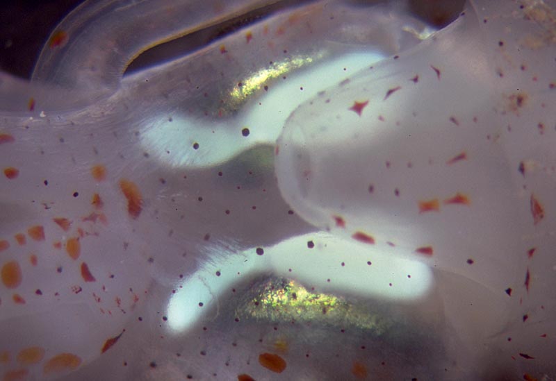
Figure. Ventral views of Onychoteuthis sp. showing photophores. Left - Arrows indicate visceral photophores (ocular photophores also visible), juvenile. Right - Ocular photophores of a different squid. Both squid from Hawaii waters. Photographs by R. Young.
Species of Onychoteuthis are commonly seen in surface waters at night and are often collected by dipnet at nightlight stations. Only young squid are normally captured in standard midwater trawls; apparently older squids avoid the trawls. One non-standard method of collection results from individuals being able to leap high out of the water and, sometimes, land on the deck of a ship. The type specimens of O. bergii were collected in this way: 'Our two specimens came to us as part of the rich inheritance of specimens from our brave Bergius, who died in the Cape of Good Hope in January of this year of tuberculosis, the victim of his own tireless efforts at collecting and observing plants and animals. His journal from the voyage reveals that these specimens flew on board the ship one night. One was found the following morning on the foredeck, the other in the crow’s nest, thirty feet above the sea surface, more evidence to support Aristotle’s remarks on their power of flight. The very elastic lateral flaps or fins may be of particular use in this respect. This occurred in May, 1816, approximately 100 miles west of the Cape. I have heard from other seafarers that other similar animals were collected in this way at about the same time.' (Translated from Lichtenstein 1818.)
Brief diagnosis:
An onychoteuthid ...
- with photophores (unique character).
- with gladius visible beneath skin in dorsal midline.
- with 8-13 occipital folds (three primary/ventrolateral and 5-8 secondary/dorsolateral).
Characteristics
- Arms
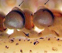
- Arm suckers with distal, fleshy thickening (see arrows in photograph below) in most species. This delicate thickening or knob is not obvious on all suckers and when present it is easily damaged during capture.
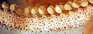
- Protective membranes low and with trabeculae fused to sucker base and not clearly seen as separate entities (see photograph below).
- Arm suckers with distal, fleshy thickening (see arrows in photograph below) in most species. This delicate thickening or knob is not obvious on all suckers and when present it is easily damaged during capture.
- Tentacular club
- Marginal suckers absent in subadults except for O. meridiopacifica (several marginal suckers present).
- Distal suckers on club restricted to terminal pad.
- Dorsal hook-series with hooks that are small in size in mid-series (see arrows on drawing below) with larger hooks proximally and distally.
- Head
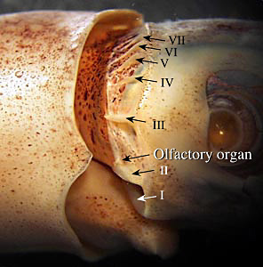
- 8-13 occipital folds present on each side of head. Arrows in the photograph below indicate the ventral 7 occipital folds. Additional very small dorsal folds are out of focus and not visible in the picture. The olfactory organ is at the posterodorsal end of occipital fold II.
- Funnel groove
- Funnel groove with distinct ridges along margin; margins convex near anterior, pointed end of groove (squid must be in good condition to see this feature).
- Mantle
- Mantle skin smooth.
- Mantle skin smooth.
- Photophores
- Large organ present ventrally on each eye (see Introduction).
- Two organs present on intestine.
- Gladius
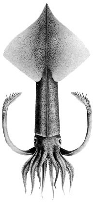
- The dorsal mantle musculature and fins are bisected by the gladius. (The resulting line down dorsal midline generally is visible through the skin - see title drawing).
- Rostrum thin (i.e., laterally compressed) and pointed (except in O. meridiopacifica).
Species comparisons
(Modified from Bolstad 2010)
| Species | Visceral photophores | Ocular photophore(s) on each eye | Club length (% ML) & hooks | Spike on distal-most ventral hooks | Habitat |
|---|---|---|---|---|---|
| O. bergii | Large, circular, posterior > anterior | One | ~25%21–23 | Present by ML 92 mm | Southern Atlantic, Indian Oceans |
| O. banksii | Large, circular, posterior >> anterior | One | ~27%20–23 | Absent | Central & North Atlantic, Gulf of Mexico |
| O. aequimanus | Large, circular, posterior > anterior | One | ~28%19–23 | May be present from ML 90 mm | Pacific Ocean south of 30°N |
| O. mollis | Large, circular, posterior >> anterior | One | Unknown | Unknown | Mediterranean (one specimen known) |
| O. compacta | Large, circular, posterior >> anterior | One | ~21%20–23 | Present by ML 60 mm | Central Pacific, and 0–40°N in western Atlantic |
| O. borealijaponica | Slender, tear-drop-shaped | One | ~27%23–27 | Present (small) | North Pacific |
| O. meridiopacifica | Minute, circular, posterior > anterior | One | ~ 19%15–19 | Absent | Indian and Southwest Pacific Oceans |
| O. lacrima | Posterior tear-drop-shaped, anterior circular, minute | One | ~ 29%20–24 | Present by ML 50 mm | North-central Pacific |
| O. prolata | Large, circular, posterior >> anterior | One | ~36%20–23 | Absent | Worldwide in temperate and tropical oceans |
| O. horstkottei | Large, circular, posterior and anterior ~subequal | Two | ~24%20–22 | Present by ML 54 mm | Eastern equatorial Pacific |
Discussion of Phylogenetic Relationships

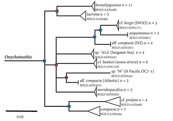
Figure. Relationships within the genus Onychoteuthis, excerpt from a combined phylogeny of onychoteuthid specimens sequenced by Bolstad et al. (2018) and previously published sequences for COI and 16S rRNA. Upper hemisphere at nodes indicates Bayesian posterior probabilities (blue > 0.95, red≤0.95); lower hemisphere indicates bootstrap support from the maximum-likelihood analysis, based on 1000 bootstrap replicates (blue > 70; red≤70). Nodes with bootstrap support below 50% have been collapsed. Barcode Index Numbers (BINs) for COI are indicated. © 2018 K.S.R. Bolstad Modified from Fig. 3 in Bolstad et al. (2018).
A recent mitochondrial assessment of the family Onychoteuthidae (Bolstad et al., 2018) brought several interesting insights into the genus Onychoteuthis. Several undescribed taxa were revealed (species with letter names in the phylogeny above, e.g. 'sp. W'). Some of these, e.g., O. compacta, O. aff. compacta (Atlantic), and O. aff. compacta (NZ) share morphological characters that were formerly believed to be species specific, such as the absence of chromatophores from a large patch of the posterior ventral mantle epidermis. These three species appear to have developed this character independently, as they all fall within different sub-clades, dispersed among taxa that do not share this character. Further study is needed in order to morphologically differentiate the additional 'compacta-like' taxa so that they can be formally described.

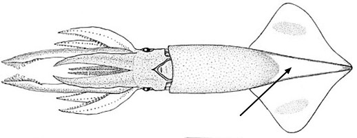
Figure. Arrow indicates region on posterior ventral mantle where chromatophores are absent in Onychoteuthis 'compacta-like' species. © K.S.R. Bolstad
Behavior
Paralarvae of Onychoteuthis, like the paralarvae of many other squid, have the ability to withdraw their head and arms into their mantle cavity. This behavior is, presumably, defensive.
Life History
Most paralarval Onychoteuthis are easily recognized by the pointed rostrum on the gladius. If the fins are damaged, the rostrum projects well beyond the fins (see photographs of the paralarvae in the previous section). The photograph on the left (ventral view, posterior end of a paralarva) shows the rostrum barely emerging and the photograph on the right (side view, posterior end of a paralarva) shows the rostrum just reaching to the edge of the fins. Even if the rostrum is not protruding due to damage, it still can be easily seen.

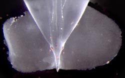
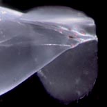
Figure. Ventral and side views of posterior ends of Onychoteuthis sp. paralarva, off Hawaii. Photographs by R. Young.
The adult female looses its tentacles apparently near the time of spawning. Spent females which are very flaccid were originally described as belonging to the genus Chaunoteuthis. In the photograph below the upper white arrow points to a scar that parallels much of the sissors cut through the mantle. This scar was apparently made by the male when mating. The lower arrow points to one of about 9 spermatangia embedded in the mantle muscle that apparently entered at the scar.

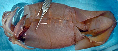
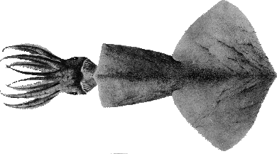
Figure. Left - Ventral view, female "Chaunoteuthis stage" of Onychoteuthis sp. , a mated and presumably (i.e. damage prevented assessment) spent female, preserved, 155 mm ML. Photograph by R. Young. Right - Dorsal view of "Chaunoteuthis mollis". Drawing from Pfeffer (1912).
Distribution
Species of Onychoteuthis are absent from the Arctic, upper boreal Atlantic, subantarctic and Antarctic waters, but occur in all other oceans (Nesis, 1982/87).
References
Bolstad, K.S.R. 2010. Systematics of the Onychoteuthidae Gray, 1847 (Cephalopoda: Oegopsida). Zootaxa, 2696: 186pp.
Bolstad, K.S.R., Heather E. Braid, Jan M. Strugnell, Annie R. Lindgren, Alexandra Lischka, Tsunemi Kubodera, Vladimir L. Laptikhovsky, & Alvaro Roura Labiaga A mitochondrial phylogeny of the family Onychoteuthidae Gray, 1847 (Cephalopoda: Oegopsida) Molecular Phylogenetics and Evolution 2018-08-07;128(N/A):88-97.
Lichtenstein, H.C. 1818. Onychoteuthis, Sepien mit Krallen. Isis, 1818: 1591–1592.
Kubodera, T., U. Piakowski, T. Okutani and M. R. Clarke. 1998. Taxonomy and zoogeography of the family Onychoteuthidae. Smiths. Contr. to Zoology, No. 585: 277-291.
Nesis, K. N. 1982/87. Abridged key to the cephalopod mollusks of the world's ocean. 385+ii pp. Light and Food Industry Publishing House, Moscow. (In Russian.). Translated into English by B. S. Levitov, ed. by L. A. Burgess (1987), Cephalopods of the world. T. F. H. Publications, Neptune City, NJ, 351pp.
Young, R. E. 1972. The systematics and areal distribution of pelagic cephalopods from the seas off Southern California. Smithson. Contr. Zool., 97: 1-159.
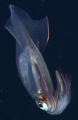
Title Illustrations

| Scientific Name | Onychoteuthis borealijaponica |
|---|---|
| Location | Monterey Bay Canyon, Northeast Pacific at 36.7°N, 122.1°W. |
| Comments | Image courtesy of the Monterey Bay Aquarium Research Institute (MBARI). You must obtain permission from MBARI to use this photo; please contact pressroom@mbari.org for further information |
| Acknowledgements | Susan Von Thun, photo editing, MBARI |
| Specimen Condition | Live Specimen |
| View | Side |
| Copyright | © 2011 MBARI |
About This Page
K.S.R. Bolstad

Auckland University of Technology
Michael Vecchione

National Museum of Natural History, Washington, D. C. , USA
Richard E. Young

University of Hawaii, Honolulu, HI, USA
Kotaro Tsuchiya

Tokyo University of Fisheries, Tokyo, Japan
Correspondence regarding this page should be directed to K.S.R. Bolstad at
kbolstad@aut.ac.nz
Page copyright © 2019 K.S.R. Bolstad, Michael Vecchione , Richard E. Young , and Kotaro Tsuchiya
 Page: Tree of Life
Onychoteuthis .
Authored by
K.S.R. Bolstad, Michael Vecchione, Richard E. Young, and Kotaro Tsuchiya.
The TEXT of this page is licensed under the
Creative Commons Attribution-NonCommercial License - Version 3.0. Note that images and other media
featured on this page are each governed by their own license, and they may or may not be available
for reuse. Click on an image or a media link to access the media data window, which provides the
relevant licensing information. For the general terms and conditions of ToL material reuse and
redistribution, please see the Tree of Life Copyright
Policies.
- Content changed 26 March 2019
Citing this page:
Bolstad, K.S.R., Michael Vecchione, Richard E. Young, and Kotaro Tsuchiya. 2019. Onychoteuthis . Version 26 March 2019 (under construction). http://tolweb.org/Onychoteuthis/19955/2019.03.26 in The Tree of Life Web Project, http://tolweb.org/





 Go to quick links
Go to quick search
Go to navigation for this section of the ToL site
Go to detailed links for the ToL site