Asperoteuthis lui
Heather Braid, Richard E. Young, and Michael VecchioneIntroduction
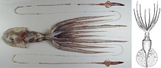
Asperoteuthis lui is a large chiroteuthid. The largest known specimen has a mantle length of 363 mm. The tentacles of this species are long and slender, with the longest intact tentacles extending 2065 mm. This species is known from a small number of specimens, none of which were fully intact and the smallest was 175 mm ML (however, see "Nesis's new chiroteuthid species"). It appears to have a circumglobal distribution in the high latitudes of the Southern hemisphere.
Brief diagnosis:
An Asperoteuthis ...
- Club with >> 100 suckers.
- Club suckers with 5-7 sharp, conical teeth.
Characteristics
- Arms
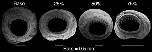
- Suckers with ~ 7-10 teeth on distal half of inner ring; proximal half smooth.
- Sucker-teeth with variable shape depending on sucker position on arm; suckers at arm base with broad teeth with blunt and rounded tips; sucker teeth become more slender and pointed with more distal position on arm; suckers in ~ distal quarter of arm with conical, sharply pointed teeth.
- Tentacle Club
- Suckerless proximal portion of club bearing mostly fused trabeculae on expanded protective menbranes ~ 25% of club length, distal sucker-bearing portion with short expanded protective membranes ~ 75% of CL.
- Proximal section of club ~ 1/3 of club length.
- Distal portion of club with ~ 120-180 suckers.
- Club suckers in 4 longitudinal series (See * under Comments).
- Tentacle-club suckers
- Inner ring with ~ 5-7 sharp, conical teeth on distal half; proximal half smooth.
 Click on an image to view larger version & data in a new window
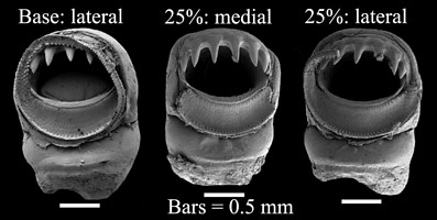
Figure. Club-sucker dentition of A. lui, NIWA 93268, no ML, 79 mm CL. Left - Lateral sucker from basal region of club showing structure of outer sucker ring. Medial - Medial sucker from ~ 25% position on club. Right - Lateral sucker, also from the 25% position. Note that lateral and medial suckers are about the same diameter. Scanning electron micrographs from Braid (2016).
- Inner ring with ~ 5-7 sharp, conical teeth on distal half; proximal half smooth.
- Tentacle photophores
- Midline of aboral surface of club with five smaller proximal and two larger distal photophores; the lateral two, at least, have aboral openings.
- Lateral margins of aboral surface of club core with small, embedded photophores; about 11-16 on dorsal margin, about 9-12 on ventral margin.
- Tentacular stalk with ~ 120 knob-like photophores that alternate in size; ~ distal 5% of stalk with only small photophores.
 Click on an image to view larger version & data in a new window

Figure. Aboral views of the tentacular club of A. lui; same club and credits as above.
- Midline of aboral surface of club with five smaller proximal and two larger distal photophores; the lateral two, at least, have aboral openings.
- Head
- Beaks: Lower mandible
- Lateral wall fold present.
- Height length nearly equals baseline length.
- Free crest length shorter than hood length.
- Beaks: Upper mandible
- Shoulder step present by LRL 3.3 in corresponding lower mandible.
- Shoulder step present by LRL 3.3 in corresponding lower mandible.
- Eye Photophores
- Single elongate photophore on ventral surface of each eye.
- Single elongate photophore on ventral surface of each eye.
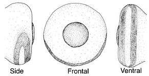
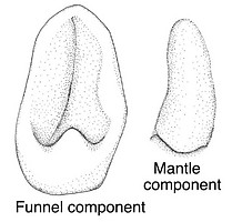
- Funnel-mantle locking-apparatus
- Funnel component approximately ear shaped; tragus long, low,
antitragus well developod. - Mantle component oval, posteriorly undercut.
- Funnel component approximately ear shaped; tragus long, low,
- Beaks: Lower mandible
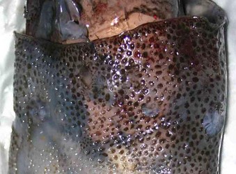
- Mantle
- Mantle covered with numerous, circular depressions containing more darkly pigmented skin.
- Skin covered with tiny (0.1-0.2 mm high) tubercles (not visible in photograph below).
- Fins
- Fins circular in combined outline.
- Fins circular in combined outline.
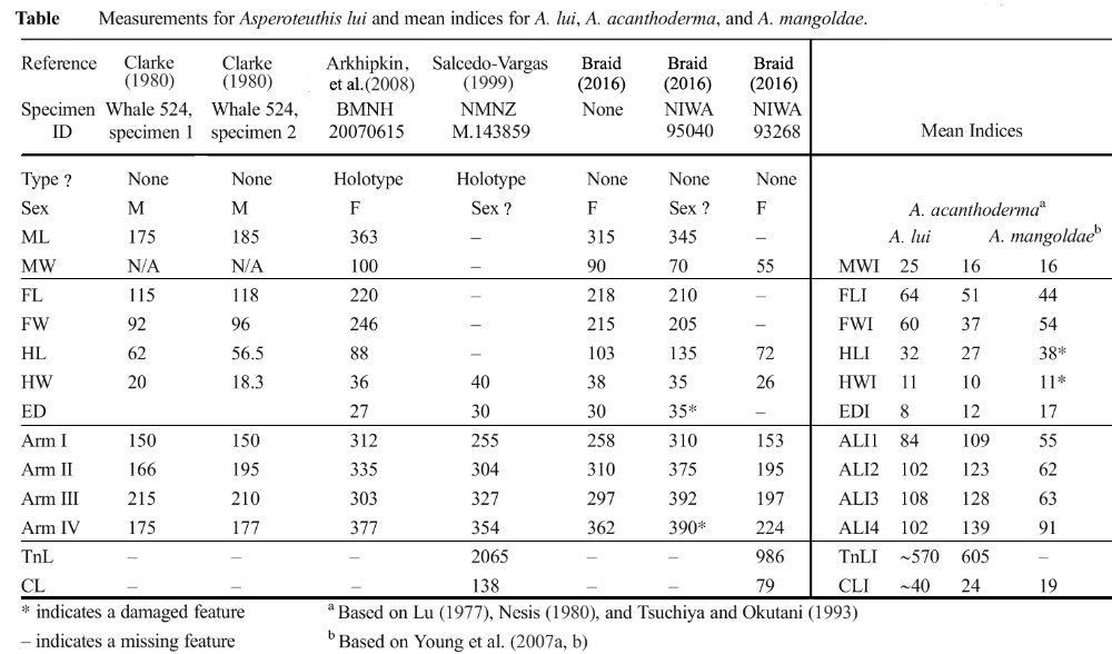
- Measurements
Comments
In contrast to what is given above, the type description described the lateral club suckers as being larger than the middle ones; this was presumably due to damage to the type (e.g., sucker rings were absent) which was found in the stomach of a fish. This and other minor differences are discussed in Braid (2016).
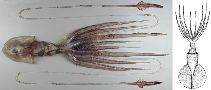
*Both the club drawing (above), and the photograph on which it is based, seem to have more than 4 series of suckers. Presumably this is a result of the club's ability to contract and extend. Typically the oegopsid club has limited ability is this regard and there is little confusion over the number of club sucker-series. The clubs of the chiroteuthid-families may be an exception to this as they are derived from the highly contractile/extensile tentacular stalk rather than from a tentacular club (see the chiroteuthid families page).
The above description was taken mostly from Braid (2016); see that paper for a more complete coverage of this species.
Nomenclature
Braid (2016) demonstrated that Asperoteuthis nesisi Arkhipkin & Laptikhovsky, 2008 (see notes from the type description of A. nesisi here), and Clarke, 1980 "?Mastigoteuthis A" (see notes from Clarke's description here) belong in Asperoteuthis lui (see notes from the type description of A. lui here). Her evidence was based on both morphological and molecular data.
The status of Nesis's two large paralarvae reported from the southwestern Atlantic as Chiroteuthis sp. n. and later (1982/87) as n. gen. B, which may belong to A. lui, is still unresolved. Nesis's more detailed publication of these paralavae can be seen here.
Discussion of Phylogenetic Relationships
- Molecular analysis
- DNA data from mitochondrial genes COI, 16S rRNA, and 12S rRNA was analyzed From squid identified as '?Mastigoteuthis A' and A. lui and compared with known data from the type specimen of A. nesisi and A. mangoldae (the outgroup) to produce the following tree using maximum-likehood software.
- DNA data from mitochondrial genes COI, 16S rRNA, and 12S rRNA was analyzed From squid identified as '?Mastigoteuthis A' and A. lui and compared with known data from the type specimen of A. nesisi and A. mangoldae (the outgroup) to produce the following tree using maximum-likehood software.

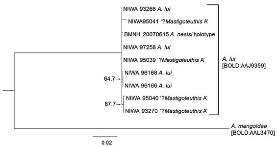
Fig. Combined maximum likelihood phylogeny of specimens now placed in A. lui; modified from Braid (2016).
For the A. lui clade, divergences for all specimens and all genes ranged from 0 to 1.5%. The divergence between this clade and the outgroup divergences ranged from 5.4 to 20.9 with divergences for COI, at 19.0% to 20.9%, being much greater those of the other genes.
Distribution
Type locality - Cook Straight, New Zealand. Taken from the stomach of a ling (fish).
General distribution - A. lui is known from the Pacific Ocean, New Zealand waters, and from the South Atlantic, near the Falkland Islands. These localities suggest that it may have a circumpolar distribution in the Southern Ocean (Braid, 2016).
References
Arkhipkin, A. I. and V. Laptikhovsky. 2008. Discovery of the fourth species of the enigmatic chiroteuthid squid Asperoteuthis (Cephalopoda: Oegopsida) and extension of the range of the genus to the South Atlantic. Journal of Molluscan Studies, 74: 203-207.
Braid, H.E. 2016. Resolving the taxonomic status of Asperoteuthis lui Salcedo-Vargas, 1999 (Cephalopoda, Chiroteuthidae) using integrative taxonomy. Mar. Biodiv. DOI 10.1007/s12526-016-0547-5
Nesis, K. N. 1974. Oceanic cephalopods of the southwestern Atlantic Ocean. Trudy Inst. Okean. Shirshova Akad. Nauk SSSR, 98: 51-75.
Nesis, K. N. 1982/87. Abridged key to the cephalopod mollusks of the world's ocean. 385,ii pp. Light and Food Industry Publishing House, Moscow. (In Russian.). Translated into English by B. S. Levitov, ed. by L. A. Burgess (1987), Cephalopods of the world. T. F. H. Publications, Neptune City, NJ, 351pp.
Salcedo-Vargas, M.A. 1999. An asperoteuthid squid (Mollusca: Cephalopoda: Chiroteuthidae) from New Zealand misidentified as Architeuthis. Mitteilungen aus dem Museum fur Naturkunde in Berlin, Zoologische Reihe 75:47-49.
Title Illustrations

| Scientific Name | Asperoteuthis lui |
|---|---|
| Location | Just east of Photograph: Chatham Isl., New Zealand at 43.79°S, 174.54°W, 810–811 m trawl depth. Drawing: Off Falkland Islands at 53°44’S 58°46’W |
| Acknowledgements | National Institute of Water & Atmospheric Research (NIWA). The title drawing is from: Arkhipkin, A. I. and V. Laptikhovsky. 2008. Discovery of the fourth species of the enigmatic chiroteuthid squid Asperoteuthis (Cephalopoda: Oegopsida) and extension of the range of the genus to the South Atlantic. Journal of Molluscan Studies, 74: 203-207. |
| Sex | Female |
| View | Ventral |
| Size | Fin missing, ML unknown; 79 mm CL, 5.4 mm LRL |
| Collection | Photograph: NIWA 93268; Drawing: BMNH 20070615 |
| Type | Drawing: Holotype of A. nesisi |
| Copyright | © Darren Stevens |
About This Page
Heather Braid

Auckland University of Technology
Richard E. Young

University of Hawaii, Honolulu, HI, USA
Michael Vecchione

National Museum of Natural History, Washington, D. C. , USA
Correspondence regarding this page should be directed to Heather Braid at
heather.braid@gmail.com
Page copyright © 2019 Heather Braid, Richard E. Young , and Michael Vecchione
 Page: Tree of Life
Asperoteuthis lui .
Authored by
Heather Braid, Richard E. Young, and Michael Vecchione.
The TEXT of this page is licensed under the
Creative Commons Attribution-NonCommercial License - Version 3.0. Note that images and other media
featured on this page are each governed by their own license, and they may or may not be available
for reuse. Click on an image or a media link to access the media data window, which provides the
relevant licensing information. For the general terms and conditions of ToL material reuse and
redistribution, please see the Tree of Life Copyright
Policies.
- First online 15 November 2007
- Content changed 20 August 2019
Citing this page:
Braid, Heather, Richard E. Young, and Michael Vecchione. 2019. Asperoteuthis lui . Version 20 August 2019. http://tolweb.org/Asperoteuthis_lui/112030/2019.08.20 in The Tree of Life Web Project, http://tolweb.org/






 Go to quick links
Go to quick search
Go to navigation for this section of the ToL site
Go to detailed links for the ToL site